In Business Since 2004

RNA Modification Analysis

LC-MS Based RNA Modification Analysis Services
At Cassia, LLC, we continue to empower scientists with new cutting‑edge reagents and tools to explore one of the most dynamic and influential areas of RNA research. Using state‑of‑the‑art LC‑MS and LC‑MS/MS platforms, we deliver highly sensitive, precise, and accurate detection of nucleoside modifications in mRNA, rRNA, tRNA, and custom RNA samples. Our systems (with 60-80% RNA coverage) can readily identify common modifications — such as acetylation, prenylation, methylation, and pseudouridinylation — and can be customized to analyze additional targets based on project needs. RNA modifications play essential roles in RNA quality control, translation, stability, and cellular stress response, and their dysregulation is linked to numerous human diseases, including cancer, neurological disorders, and mitochondrial syndromes. Cassia’s mass spectrometry services allows researchers to explore these mechanisms with confidence, offering comprehensive qualitative and quantitative insights into RNA modification landscapes.
- mRNA Modification Analysis: High‑resolution mapping and quantitation of key epigenetic RNA marks, including m6A, m6Am, m7G, Nm, and m5C. These modifications regulate mRNA splicing, transport, stability, and translation and are controlled by enzyme systems closely linked to human health, disease, and RNA‑based therapeutics.
- rRNA Modification Analysis: Advanced analysis supporting research in ribosome biology, translational regulation, and ribosomal dysfunction. rRNA contains dense patterns of 2′‑O‑methylation, pseudouridylation, and other essential modifications that ensure ribosome assembly, stability, and translational fidelity. Altered rRNA modification patterns are increasingly associated with cancer, developmental disorders, and ribosomopathies.
- tRNA Modification Analysis: tRNAs are the most chemically modified RNA species, averaging ~13 modifications per molecule. These modifications are critical for codon recognition, decoding fidelity, folding, stability, and stress adaptation. Detailed tRNA modification profiles provide deep insight into gene expression regulation and disease‑associated modification shifts.
With deep technical expertise and cutting‑edge instrumentation, Cassia, LLC offers unmatched capability in RNA modification analysis. Whether you are studying molecular mechanisms, identifying biomarkers, or advancing RNA‑based technologies or therapeutics, our team provides the accuracy, insight, and responsiveness needed to accelerate discovery. Contact the experts at Cassia, LLC to learn more about our RNA analysis services.
Workflow for RNA Modification Analysis
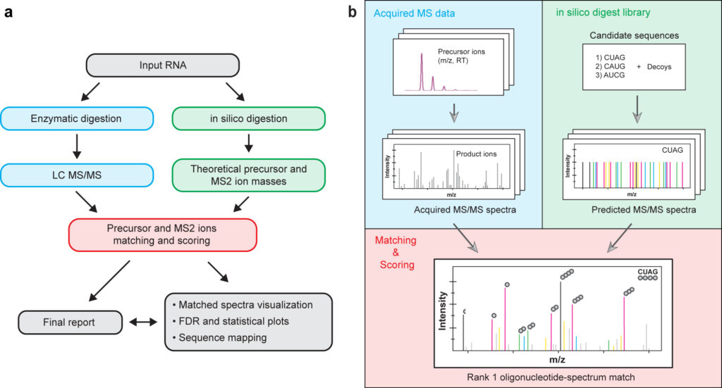
The flowchart (a) highlights the main components of a typical RNA processing and analysis workflow. While the graphical overview (b) of the database matching process. In both panels, blue shading refers to the experimental data acquisition, green shading refers to the in silico generated theoretical library and red shading refers to the matching and scoring steps.
Enzymes Utilized for Routine RNA Hydrolysis
There are several Ribonucleases (RNases) for the routine manipulation of RNA. Endoribonucleases recognize and cleave internal RNA sequences with distinct activities of each RNase — including recognition of specific sequence or structural elements and reaction products — enabling numerous experimental approaches for generating the LC-MS and LC-MS/MS datasets.

Ribonuclease A (RNAse A, EC 3.1.27.5): a robust endoribonuclease commonly used in RNA epigenetic analysis because its well‑defined cleavage specificity provides a stable biochemical baseline against which RNA modifications can be mapped. It preferentially cleaves single‑stranded RNA at the 3′ side of unmodified pyrimidines (e.g. cytidine > unidine). Certain modifications; such as 2′‑O‑methylation, pseudouridine, or base‑modified cytidines and uridines, often exhibit altered or reduced RNAse A digestion activities. By comparing RNAse A cleavage profiles with and without these modifications, cleavage-resistant regions can be indentified, thus helping to localize and characterize epitranscriptomic marks without relying on sequence changes alone.
Ribonuclease 4 (RNAse 4, EC 3.1.27.-): a highly conserved member of the RNAse A superfamily that preferentially cleaves the 3′ side of uridine‑rich RNA sequences (e.g. U/A and U/G), generating defined small RNA fragments. Although RNAse 4 is not itself an “epigenetic writer, reader, or eraser,” it is increasingly relevant to RNA epigenetic analysis because its activity can shape the RNA substrate landscape available for chemical modification profiling. Cleavage by RNAse 4 can reveal or obscure specific RNA modifications (e.g. pseudo-, N1-methylpseudo-, dihydro-, and 5-methoxyuridine species) by altering local secondary structure and fragment composition, and its products may retain modification marks that are useful for high‑resolution mapping techniques such as LC‑MS/MS.

Ribonuclease T1 (RNAse T1, EC 4.6.1.24): a guanosine-specific endoribonuclease that cleaves RNA at the 3′ side of unmodified guanosine residues, a property that makes it particularly valuable in RNA epigenetic analysis and RNA modification mapping. In LC-MS and LC-MS/MS applications, RNAse T1 generates predictable oligonucleotide fragments whose cleavage patterns are altered when guanosines carry chemical modifications (e.g. N7-methylguanosine, 2′-O-methylguanosine, or other bulky adducts such as Wybutosine), leading to missed or shifted cuts. By comparing RNAse T1 digestion profiles with complementary nucleases or across different conditions, researchers can infer the presence, location, and sometimes the stoichiometry of RNA modifications, making RNAse T1 a foundational tool in both classical and modern epitranscriptomic workflows.
Endonuclease V (Endo V, EC 3.1.21.7): a inosine-specific endoribonuclease — formed through A‑to‑I RNA editing — introducing a site‑specific cleavage at the 3′ side of inosine bases, making it a powerful biochemical tool for RNA modification mapping. Because inosine base‑pairs like guanosine and cannot be directly distinguished by standard sequencing, Endo V–mediated cleavage enriches or flags edited sites, enabling more accurate detection of A‑to‑I editing events at single‑nucleotide resolution. In RNA epigenetic analysis workflows, Endo V is commonly used to validate editing sites, enhance detection sensitivity, and complement other nucleases mappings to construct a more detailed map of RNA editing.
RNA Modifications Identified by LC-MS/MS Analysis
| Nucleoside Name | Short Name | RNAMods Code (2023) | Common RNA Type | Routinely Detected |
|---|---|---|---|---|
| Adenosine | A | A | mRNA, rRNA, snRNA, snoRNA, tRNA | Yes |
| 1-Methyladenosine | m1A | Ѣ | rRNA, tRNA | Yes |
| 1-Methyl-2'-O-Methyladenosine | m1Am | œ | mRNA | Yes |
| 2-Methyladenosine | m2A | ɿ | rRNA, tRNA | Yes |
| 2-Methylthio-N6-(cis-Hydroxyisopentenyl)adenosine | ms2io6A | ≠ | tRNA | Yes |
| 2-Methylthio-N6-Hydroxynorvalylcarbamoyladenosine | ms2hn6A | ≈ | tRNA | Yes |
| 2-Methylthio-N6-Isopenenyladenosine | ms2i6A | * | tRNA | Yes |
| 2-Methylthio-N6-Methyladenosine | ms2m6A | ∞ | tRNA | Yes |
| 2-Methylthio-N6-Threonylcarbamoyladenosine | ms2t6A | [ | tRNA | Yes |
| N6-Acetyladenosine | ac6A | ⇓ | tRNA | No |
| N6-(cis-Hydroxyisopentenyl)adenosine | io6A | Ỽ | tRNA | Yes |
| N6-Hydroxynorvalylcarbamoyladenosine | hn6A | √ | tRNA | Yes |
| N6-Glycinylcarbamoyladenosine | g6A | ≡ | tRNA | Yes |
| N6-Isopentenyladenosine | i6A | Ч | tRNA | Yes |
| N6-Methyladenosine | m6A | Ж | mRNA, rRNA, snRNA, tRNA | Yes |
| N6-Threonylcarbamoyladenosine | t6A | 6 | tRNA | Yes |
| N6-Methyl-N6-Threonylcarbamoyladenosine | m6t6A | E | tRNA | Yes |
| N6-Methyl-2'-O-Methyladenosine | m6Am | χ | mRNA, snRNA | Yes |
| N6,N6-Dimethyladenosine | m6,6A | ζ | rRNA | Yes |
| N6,N6-Dimethyl-2'-O-Methyladenosine | m6,6Am | η | rRNA | Yes |
| 2'-Deoxyadenosine | dA | unassigned | DNA, Chimeric Oligonucleotide | No |
| 2'-O-Methyladenosine | Am | ʍ | rRNA, snRNA, snoRNA, tRNA | Yes |
| 2'-O-Ribosyladenosine (Phosphate) | Ar(p) | ʩ | tRNA | Yes |
If you don’t find the exact modification you’re looking for, please do not hesitate to ask! We are here to help and can quickly modify our protocols to identify additional nucleotide modifications.
| Nucleoside Name | Short Name | RNAMods Code (2023) | Common RNA Type | Routinely Detected |
|---|---|---|---|---|
| Cytidine | C | C | mRNA, rRNA, snRNA, snoRNA, tRNA | Yes |
| 2-Lysidine | k2C | } | tRNA | Yes |
| 2-Thiocytidine | s2C | ʤ | tRNA | Yes |
| 3-Methylcytidine | m3C | Щ | tRNA | Yes |
| 5-Formylcytidine | f5C | > | tRNA | Yes |
| 5-Formyl-2'-O-Methylcytidine | f5Cm | ° | tRNA | Yes |
| 5-Hydroxycytidine | ho5C | Ç | rRNA | Yes |
| 5-Hydroxymethylcytidine | hm5C | Ƣ | tRNA | Yes |
| 5-Methylcytidine | m5C | ? | mRNA, rRNA, tRNA | Yes |
| 5-Methyl-2'-O-Methylcytidine | m5Cm | τ | rRNA, tRNA | Yes |
| N4-Acetylcytidine | ac4C | M | rRNA, tRNA | Yes |
| N4-Acetyl-2'-O-Methylcytidine | ac4Cm | ℵ | rRNA, tRNA | Yes |
| N4-Methylcytidine | m4C | ν | rRNA | Yes |
| N4-Methyl-2'-O-Methylcytidine | m4Cm | λ | rRNA | Yes |
| N4,N4-Dimethyl-2'-O-Methylcytidine | m4,4Cm | β | rRNA | Yes |
| 2'-Deoxycytidine | dC | unassigned | DNA, Chimeric Oligonucleotide | No |
| 2'-O-Methylcytidine | Cm | B | rRNA, snRNA, tRNA | Yes |
If you don’t find the exact modification you’re looking for, please do not hesitate to ask! We are here to help and can quickly modify our protocols to identify additional nucleotide modifications.
| Nucleoside Name | Short Name | RNAMods Code (2023) | Common RNA Type | Routinely Detected |
|---|---|---|---|---|
| Guanosine | G | G | mRNA, rRNA, snRNA, snoRNA, tRNA | Yes |
| 1-Methylguanosine | m1G | K | rRNA, tRNA | Yes |
| 1-Methyl-2'-O-Methylguanosine | m1Gm | ε | tRNA | No |
| 7-Aminomethyl-7-Deazaguanosine | preQ1 | ∉ | tRNA | Yes |
| 7-Cyano-7-Deazaguanosine | preQ0 | φ | tRNA | Yes |
| 7-Methylguanosine | m7G | 7 | mRNA, rRNA, tRNA | Yes |
| N2-Methylguanosine | m2G | L | rRNA, snRNA, tRNA | Yes |
| N2-Methyl-2'-O-Methylguanosine | m2Gm | γ | rRNA, tRNA | Yes |
| N2-Methyl-7-Methylguanosine | m2,7G | ∨ | snRNA, snoRNA | Yes |
| N2-Methyl-7-Methyl-2'-O-Methylguanosine | m2,7Gm | æ | tRNA | Yes |
| N2,N2-Dimethylguanosine | m2,2G | R | rRNA, tRNA | Yes |
| N2,N2-Dimethyl-7-Methylguanosine | m2,2,7G | ∠ | snRNA, snoRNA | Yes |
| N2,N2-Dimethyl-2'-O-Methylguanosine | m2,2Gm | | | rRNA, tRNA | Yes |
| 2'-Deoxyguanosine | dG | unassigned | DNA, Chimeric Oligonucleotide | No |
| 2'-O-Methylguanosine | Gm | # | rRNA, snRNA, tRNA | Yes |
| 2'-O-Ribosylguanosine (Phosphate) | Gr(p) | ℑ | tRNA | Yes |
| Archaeosine | G+ | ( | tRNA | Yes |
| Epoxyqueuosine | oQ | ς | tRNA | Yes |
| Hydroxywybutosine | OHyW | ⊆ | tRNA | Yes |
| Undermodified Hydroxywybutosine | OHyWx | š | tRNA | Yes |
| Isowyosine | imG2 | ⊇ | tRNA | Yes |
| Queuosine | Q | Q | tRNA | Yes |
| 5''-β-(D)-Galactosylqueuosine | galQ | 9 | tRNA | Yes |
| 5''-β-(D)-Mannosylqueuosine | manQ | 8 | tRNA | Yes |
| Wybutosine | yW | Y | rRNA, tRNA | Yes |
| 2-Peroxywybutosine | o2yW | W | tRNA | Yes |
| Wyosine | imG | € | tRNA | Yes |
| 4-Demethylwyosine | imG-14 | † | tRNA | Yes |
| 7-Methylwyosine | mimG | ∑ | tRNA | Yes |
If you don’t find the exact modification you’re looking for, please do not hesitate to ask! We are here to help and can quickly modify our protocols to identify additional nucleotide modifications.
| Nucleoside Name | Short Name | RNAMods Code (2023) | Common RNA Type | Routinely Detected |
|---|---|---|---|---|
| Inosine | I | I | tRNA | Yes |
| N1-Methylinosine | m1I | O | tRNA | Yes |
| N1-Methyl-2'-O-Methylinosine | m1Im | ξ | tRNA | No |
| 2'-O-Methylinosine | Im | Ш | sncRNA, tRNA | No |
If you don’t find the exact modification you’re looking for, please do not hesitate to ask! We are here to help and can quickly modify our protocols to identify additional nucleotide modifications.
| Nucleoside Name | Short Name | RNAMods Code (2023) | Common RNA Type | Routinely Detected |
|---|---|---|---|---|
| Uridine | U | U | mRNA, rRNA, snRNA, snoRNA, tRNA | Yes |
| 2-Thiouridine | s2U | 2 | tRNA | Yes |
| 2-Thio-2'-O-Methyluridine | s2Um | ∏ | tRNA | Yes |
| 3-(3-Amino-3-Carboxypropyl)uridine | acp3U | X | rRNA, tRNA | Yes |
| 3-Methyluridine | m3U | δ | rRNA | Yes |
| 3-Methyl-2'-O-Methyluridine | m3Um | σ | mRNA, rRNA | Yes |
| 4-Thiouridine | s4U | 4 | tRNA | Yes |
| 5-Aminomethyl-2-Thiouridine | nm5s2U | ∫ | tRNA | Yes |
| 5-Carbamoylmethyluridine | ncm5U | & | tRNA | Yes |
| 5-Carbamoylmethyl-2'-O-Methyluridine | ncm5Um | ~ | tRNA | Yes |
| 5-(Carboxymethoxy)uridine | cmo5U | V | tRNA | Yes |
| 5-(Methoxycarbonylmethoxy)uridine | mcmo5U | υ | tRNA | Yes |
| 5-Carboxylmethylaminomethyluridine | cmnm5U | ! | tRNA | Yes |
| 5-Carboxylmethylaminomethyl-2-Thiouridine | cmnm5s2U | $ | tRNA | Yes |
| 5-Carboxylmethylaminomethyl-2'-O-Methyluridine | cmnm5Um | ) | tRNA | Yes |
| 5-Carboxylmethyluridine | cm5U | ◊ | tRNA | Yes |
| 5-Hydroxyuridine | ho5U | ∝ | tRNA | Yes |
| 5-(Isopentenylaminomethyl)uridine | inm5U | ¾ | tRNA | Yes |
| 5-(Isopentenylaminomethyl)-2-Thiouridine | inm5s2U | Ɲ | tRNA | Yes |
| 5-(Isopentenylaminomethyl)-2'-O-Methyluridine | inm5s2Um | Ю | tRNA | Yes |
| 5-Methoxycarbonylmethyluridine | mcm5U | 1 | tRNA | Yes |
| 5-Methoxycarbonylmethyl-2-Thiouridine | mcm5s2U | 3 | tRNA | Yes |
| 5-Methoxycarbonylmethyl-2'-O-Methyluridine | mcm5Um | ∩ | tRNA | Yes |
| 5-Methoxyuridine | mo5U | 5 | tRNA | Yes |
| 5-Methylaminomethyluridine | mnm5U | { | tRNA | Yes |
| 5-Methylaminomethyl-2-Selenouridine | mnm5se2U | ≅ | tRNA | Yes |
| 5-Methylaminomethyl-2-Thiouridine | mnm5s2U | S | tRNA | Yes |
| 5-Methyluridine | m5U | T | rRNA, tRNA | Yes |
| 5-Methyl-2-Thiouridine | m5s2U | F | tRNA | Yes |
| 5-Methyl-2'-O-Methyluridine | m5Um | Ħ | tRNA | Yes |
| 5-Taurinomethyluridine | tm5U | ʭ | tRNA | Yes |
| 5-Taurinomethyl-2-Thiouridine | tm5s2U | ƕ | tRNA | Yes |
| 2'-Deoxyuridine | dU | unassigned | DNA, Chimeric Oligonucleotide | No |
| 2'-O-Methyluridine | Um | J | rRNA, snRNA, snoRNA, tRNA | Yes |
| Dihydrouridine | D | D | rRNA, tRNA | Yes |
| 5-Methyldihydrouridine | m5D | ρ | rRNA, tRNA | Yes |
| Pseudouridine | Y | P | rRNA, snRNA, snoRNA, tRNA | Yes |
| 1-Methylpseudouridine | m1Y | ] | rRNA, tRNA | Yes |
| 1-Methyl-3-(3-Amino-3-Carboxypropyl)pseudouridine | m1acp3Y | α | rRNA | Yes |
| 3-Methylpseudouridine | m3Y | Ƒ | rRNA | Yes |
| 2'-O-Methylpseudouridine | Ym | Z | rRNA, snRNA, tRNA | Yes |
| Thymidine | dT | unassigned | DNA, Chimeric Oligonucleotide | No |
If you don’t find the exact modification you’re looking for, please do not hesitate to ask! We are here to help and can quickly modify our protocols to identify additional nucleotide modifications.
| Nucleoside Name | Short Name | RNAMods Code (2023) | Reference NucleoBase | Routinely Detected |
|---|---|---|---|---|
| Adenosine (2'-O,C4'-cEt) | cEt-A | unassigned | A | No |
| 2-Fluoroadenosine | F2A | unassigned | A | No |
| 2-Fluoroinosine | F2I | unassigned | F2A → F2I | No |
| 5-Fluorocytidine | F5C | 㐭 | C | No |
| 5-Fluorouridine | F5U | unassigned | U | No |
| 5-Methyluridine (2'-O,C4'-cEt) | cEt-m5U | unassigned | m5U | No |
| 2'-Deoxy-2'-(R)-Fluoroadenosine | AF2 | 㐀 | A | No |
| 2'-Deoxy-2'-(R)-Fluorocytidine | CF2 | 㐄 | C | No |
| 2'-Deoxy-2'-(R)-Fluoroguanosine | GF2 | unassigned | G | No |
| 2'-Deoxy-2'-(R)-Fluorothymidine | TF2 | unassigned | T | No |
| 2'-Deoxy-2'-(R)-Fluorouridine | UF2 | 㐋 | U | No |
If you don’t find the exact modification you’re looking for, please do not hesitate to ask! We are here to help and can quickly modify our protocols to identify additional nucleotide modifications.
| Modification Name | Short Name | RNAMods Code (2023) | Reference NucleoBase | Routinely Detected |
|---|---|---|---|---|
| Amino C6 Linker | Ahex (AmC6) | unassigned | N | No |
| Cyanoethyl | CNEt | unassigned | I, Y | No |
| Phosphorothioate | Ps | Ps | N | No |
| Propargyl | Prp | unassigned | Y | No |
If you don’t find the exact modification you’re looking for, please do not hesitate to ask! We are here to help and can quickly modify our protocols to identify additional nucleotide modifications.
Literature
Complete List of Canonical Post-transcriptional Modifications in the Bacillus subtilis Ribosome and their Link to RbgA Driven Large Subunit Assembly
Nucleic Acids Res. 2024, 52, 11203-11217.

Abstract: Ribosomal RNA modifications in prokaryotes have been sporadically studied, but there is a lack of a comprehensive picture of modification sites across bacterial phylogeny. Bacillus subtilis is a preeminent model organism for gram-positive bacteria, with a well-annotated and editable genome, convenient for fundamental studies and industrial use. Yet remarkably, there has been no complete characterization of its rRNA modification inventory. By expanding modern MS tools for the discovery of RNA modifications, we found a total of 25 modification sites in 16S and 23S rRNA of B. subtilis, including the chemical identity of the modified nucleosides and their precise sequence location. Furthermore, by perturbing large subunit biogenesis using depletion of an essential factor rbgA and measuring the completion of 23S modifications in the accumulated intermediate, we provide a first look at the order of modification steps during the late stages of assembly in B. subtilis. While our work expands the knowledge of bacterial rRNA modification patterns, adding B. subtilis to the list of fully annotated species after Escherichia coli and Thermus thermophilus, in a broader context, it provides the experimental framework for discovery and functional profiling of rRNA modifications to ultimately elucidate their role in ribosome biogenesis and translation.
Tandem Mass Spectrometry Across Platforms
Analytical Chem. 2024, 96, 5478-5488.

Abstract: PubChem serves as a comprehensive repository, housing over 100 million unique chemical structures representing the breadth of our chemical knowledge across numerous fields including metabolism, pharmaceuticals, toxicology, cosmetics, agriculture, and many more. Rapid identification of these small molecules increasingly relies on electrospray ionization (ESI) paired with tandem mass spectrometry (MS/MS), particularly by comparison to genuine standard MS/MS data sets. Despite its widespread application, achieving consistency in MS/MS data across various analytical platforms remains an unaddressed concern. This study evaluated MS/MS data derived from one hundred molecular standards utilizing instruments from five manufacturers, inclusive of quadrupole time-of-flight (QTOF) and quadrupole orbitrap “exactive” (QE) mass spectrometers by Agilent (QTOF), Bruker (QTOF), SCIEX (QTOF), Waters (QTOF), and Thermo QE. We assessed fragment ion variations at multiple collisional energies (0, 10, 20, and 40 eV) using the cosine scoring algorithm for comparisons and the number of fragments observed. A parallel visual analysis of the MS/MS spectra across instruments was conducted, consistent with a standard procedure that is used to circumvent the still prevalent issue of mischaracterizations as shown for dimethyl sphingosine and C20 sphingosine. Our analysis revealed a notable consistency in MS/MS data and identifications, with fragment ions’ m/z values exhibiting the highest concordance between instrument platforms at 20 eV, the other collisional energies (0, 10, and 40 eV) were significantly lower. While moving toward a standardized ESI MS/MS protocol is required for dependable molecular characterization, our results also underscore the continued importance of corroborating MS/MS data against standards to ensure accurate identifications. Our findings suggest that ESI MS/MS manufacturers, akin to the established norms for gas chromatography mass spectrometry instruments, should standardize the collision energy at 20 eV across different instrument platforms.
Pytheas: A Software Package for the Automated Analysis of RNA Sequences and Modifications via Tandem Mass Spectrometry
Nat. Commun. 2022, 13, 2424-2436.
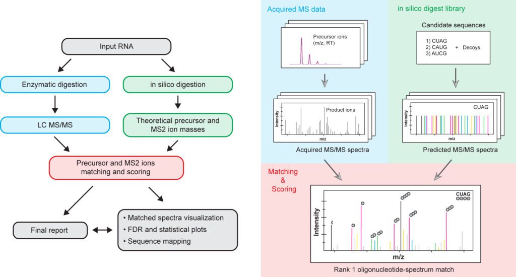
Abstract: Mass spectrometry is an important method for analysis of modified nucleosides ubiquitously present in cellular RNAs, in particular for ribosomal and transfer RNAs that play crucial roles in mRNA translation and decoding. Furthermore, modifications have effect on the lifetimes of nucleic acids in plasma and cells and are consequently incorporated into RNA therapeutics. To provide an analytical tool for sequence characterization of modified RNAs, we developed Pytheas, an open-source software package for automated analysis of tandem MS data for RNA. The main features of Pytheas are flexible handling of isotope labeling and RNA modifications, with false discovery rate statistical validation based on sequence decoys. We demonstrate bottom-up mass spectrometry characterization of diverse RNA sequences, with broad applications in the biology of stable RNAs, and quality control of RNA therapeutics and mRNA vaccines.
Quantitative Analysis of rRNA Modifications Using Stable Isotope Labeling and Mass Spectrometry
J. Am. Chem. Soc. 2014, 136, 2058-2069.
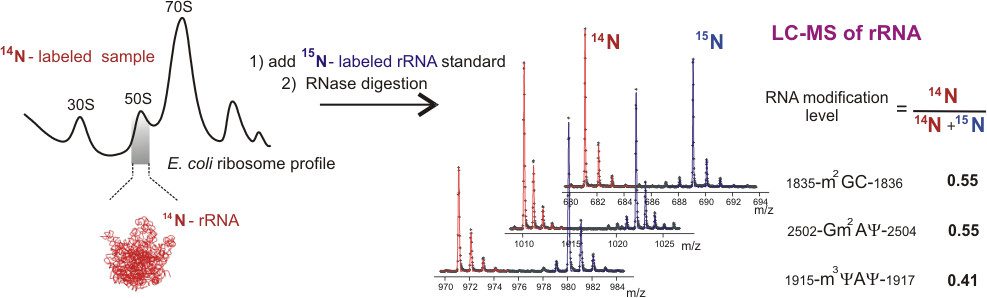
Abstract: Post-transcriptional RNA modifications that are introduced during the multistep ribosome biogenesis process are essential for protein synthesis. The current lack of a comprehensive method for a fast quantitative analysis of rRNA modifications significantly limits our understanding of how individual modification steps are coordinated during biogenesis inside the cell. Here, an LC-MS approach has been developed and successfully applied for quantitative monitoring of 29 out of 36 modified residues in the 16S and 23S rRNA from Escherichia coli. An isotope labeling strategy is described for efficient identification of ribose and base methylations, and a novel metabolic labeling approach is presented to allow identification of MS-silent pseudouridine modifications. The method was used to measure relative abundances of modified residues in incomplete ribosomal subunits compared to a mature (15)N-labeled rRNA standard, and a number of modifications in both 16S and 23S rRNA were present in substoichiometric amounts in the preribosomal particles. The RNA modification levels correlate well with previously obtained profiles for the ribosomal proteins, suggesting that RNA is modified in a schedule comparable to the association of the ribosomal proteins. Importantly, this study establishes an efficient workflow for a global monitoring of ribosomal modifications that will contribute to a better understanding of mechanisms of RNA modifications and their impact on intracellular processes in the future.
Characterization of the Ribosome Biogenesis Landscape in E. coli Using Quantitative Mass Spectrometry
J. Mol. Biol. 2013, 425, 767-779.
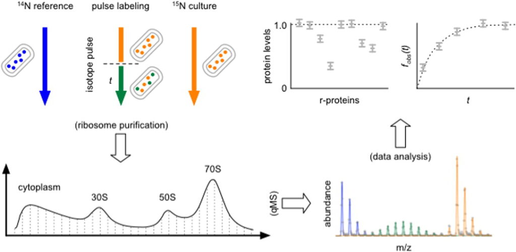
Abstract: The ribosome is an essential and highly complex biological system in all living cells. A large body of literature on the assembly of the ribosome in vitro is available, but a clear picture of this process inside the cell has yet to emerge. Here, we directly characterized in vivo ribosome assembly intermediates and associated assembly factors from wild-type Escherichia coli cells using a general quantitative mass spectrometry (qMS) approach. The presence of distinct populations of ribosome assembly intermediates was verified using an in vivo stable isotope pulse-labeling approach, and their exact ribosomal protein contents were characterized against an isotopically labeled standard. The model-free clustering analysis of the resultant protein levels for the different ribosomal particles produced four 30S assembly groups that correlate very well with previous in vitro assembly studies of the small ribosomal subunit and six 50S assembly groups that clearly define an in vivo assembly landscape for the larger ribosomal subunit. In addition, de novo proteomics identified a total of 21 known and potentially new ribosome assembly factors co-localized with various ribosomal particles. These results represent new in vivo assembly maps of the E. coli 30S and 50S subunits, and the general qMS approach should prove to be a solid platform for future studies of ribosome biogenesis across a host of model organisms.
Measuring the Dynamics of E. coli Ribosome Biogenesis Using Pulse-Labeling and Quantitative Mass Spectrometry
Mol. Biosyst. 2012, 8, 3325-3334.
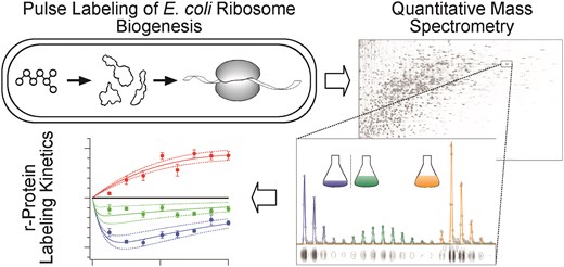
Abstract: The ribosome is an essential organelle responsible for cellular protein synthesis. Until recently, the study of ribosome assembly has been largely limited to in vitro assays, with few attempts to reconcile these results with the more complex in vivo ribosome biogenesis process. Here, we characterize the ribosome synthesis and assembly pathway for each E. coli ribosomal protein (r-protein) in vivo using a stable isotope pulse-labeling timecourse. Isotope incorporation into assembled ribosomes was measured by quantitative mass spectrometry (qMS) and fit using steady-state flux models. Most r-proteins exhibit precursor pools ranging in size from 0% to 7% of completed ribosomes, and that the sizes of these individual r-protein pools correlate well with the order of r-protein binding in vitro. Additionally, we observe anomalously large precursor pools for specific r-proteins with known extra-ribosomal functions and we have detected three r-proteins with significant turnover during steady-state growth. Taken together, this highly precise, time-dependent proteomic qMS approach should prove useful in future studies of ribosome biogenesis and could be easily extended to explore other complex biological processes in a cellular context.
Identification of 5-Hydroxycytidine at Position 2501 Concludes Characterization of Modified Nucleotides in E. coli 23S rRNA
J. Mol. Biol. 2011, 411, 529-536.
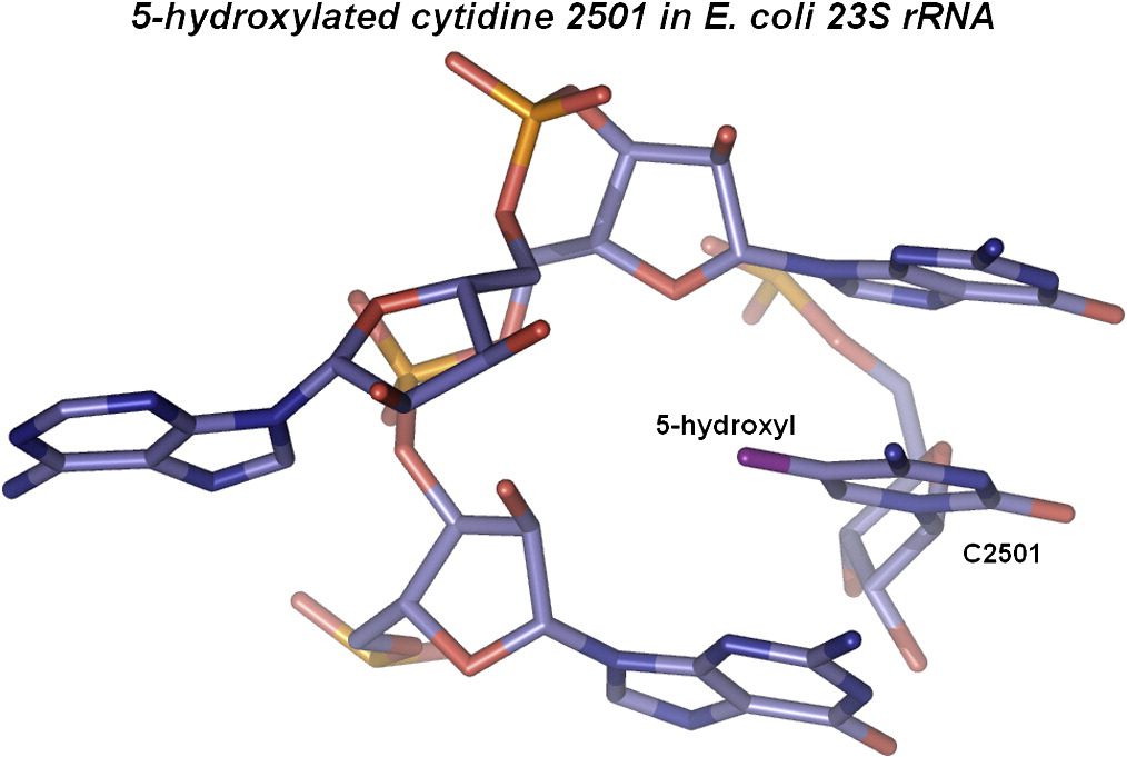
Abstract: Complete characterization of a biomolecule’s chemical structure is crucial in the full understanding of the relations between their structure and function. The dominating components in ribosomes are ribosomal RNAs (rRNAs), and the entire rRNA – but a single modified nucleoside at position 2501 in 23S rRNA – has previously been characterized in the bacterium Escherichia coli. Despite a first report nearly 20 years ago, the chemical nature of the modification at position 2501 has remained elusive, and attempts to isolate it have so far been unsuccessful. We unambiguously identify this last unknown modification as 5-hydroxycytidine – a novel modification in RNA. Identification of 5-hydroxycytidine was completed by liquid chromatography under nonoxidizing conditions using a graphitized carbon stationary phase in combination with ion trap tandem mass spectrometry and by comparing the fragmentation behavior of the natural nucleoside with that of a chemically synthesized ditto. Furthermore, we show that 5-hydroxycytidine is also present in the equivalent position of 23S rRNA from the bacterium Deinococcus radiodurans. Given the unstable nature of 5-hydroxycytidine, this modification might be found in other RNAs when applying the proper analytical conditions as described here.
A Nano-Chip-LC/MSn Based Strategy for Characterization of Modified Nucleosides Using Reduced Porous Graphitic Carbon as a Stationary Phase
J. Am. Soc. Mass Spectrom. 2011, 22, 1242-1251.
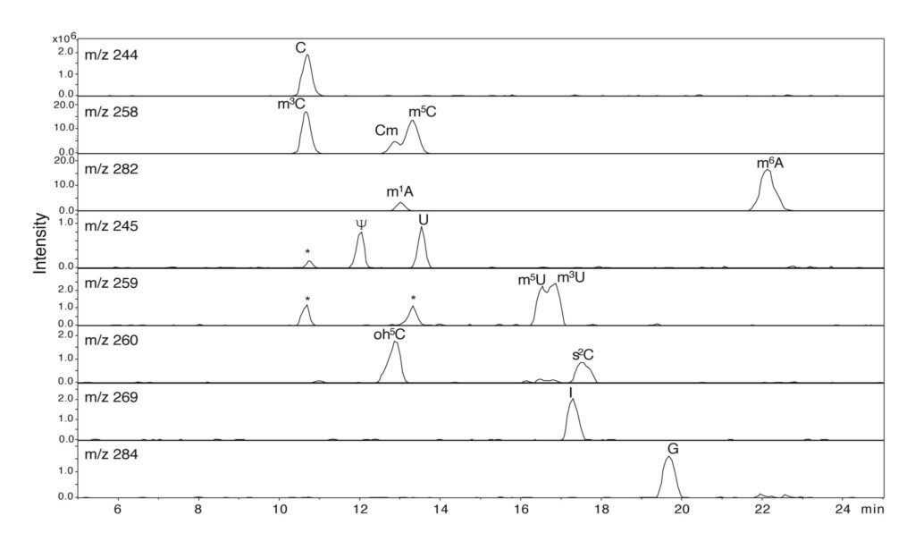
Abstract: LC/MS analysis of ribonucleosides is traditionally performed by reverse phase chromatography on silica based C18 type stationary phases using MS compatible buffers and methanol or acetonitrile gradients. Due to the hydrophilic and polar nature of nucleosides, down-scaling C18 analytical methods to a two-column nano-flow setup is inherently difficult. We present a nano-chip LC/MS ion-trap strategy for routine characterization of RNA nucleosides in the fmol range. Nucleosides were analyzed in positive ion mode by reverse phase chromatography using a 75 μ × 150 mm, 5 μ particle porous graphitic carbon (PGC) chip with an integrated 9 mm, 160 nL trapping column. Nucleosides were separated using a formic acid/acetonitrile gradient. The method was able to separate isobaric nucleosides as well as nucleosides with isotopic overlap to allow unambiguous MSn identification on a low resolution ion-trap. Synthesis of 5-hydroxycytidine (oh5C) was achieved from 5-hydroxyuracil in a novel three-step enzymatic process. When operated in its native state using formic acid/acetonitrile, PGC oxidized oh5C to its corresponding glycols and formic acid conjugates. Reduction of the PGC stationary phase was achieved by flushing the chip with 2.5 mM oxalic acid and adding 1 mM oxalic acid to the online solvents. Analyzed under reduced chromatographic conditions oh5C was readily identified by its MH+ m/z 260 and MSn fragmentation pattern. This investigation is, to our knowledge, the first instance where oxalic acid has been used as an online reducing agent for LC/MS. The method was subsequently used for complete characterization of nucleosides found in tRNAs using both PGC and C18 chips.
Stable Isotope Pulse-Chase Monitored by Quantitative Mass Spectrometry Applied to E. coli 30S Ribosome Assembly Kinetics
Methods 2009, 49, 136-141.
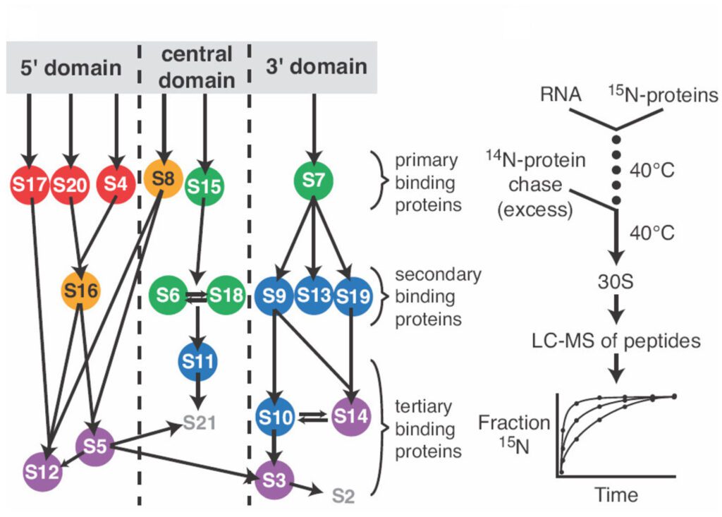
Abstract: Stable isotope mass spectrometry has become a widespread tool in quantitative biology. Pulse-chase monitored by quantitative mass spectrometry (PC/QMS) is a recently developed stable isotope approach that provides a powerful means of studying the in vitro self-assembly kinetics of macromolecular complexes. This method has been applied to the Escherichia coli 30S ribosomal subunit, but could be applied to any stable self-assembling complex that can be reconstituted from its component parts and purified from a mixture of components and complex. The binding rates of 18 out of the 20 ribosomal proteins have been measured at several temperatures using PC/QMS. Here, PC/QMS experiments on 30S ribosomal subunit assembly are described, and the potential application of the method to other complexes is discussed. A variation on the PC/QMS experiment is introduced that enables measurement of kinetic cooperativity between proteins. In addition, several related approaches to stable isotope labeling and quantitative mass spectrometry data analysis are compared and contrasted.
Quantitative Analysis of Isotope Distributions in Proteomic Mass Spectrometry Using Least-Squares Fourier Transform Convolution
Analytical Chem. 2008, 80, 4906-4917.
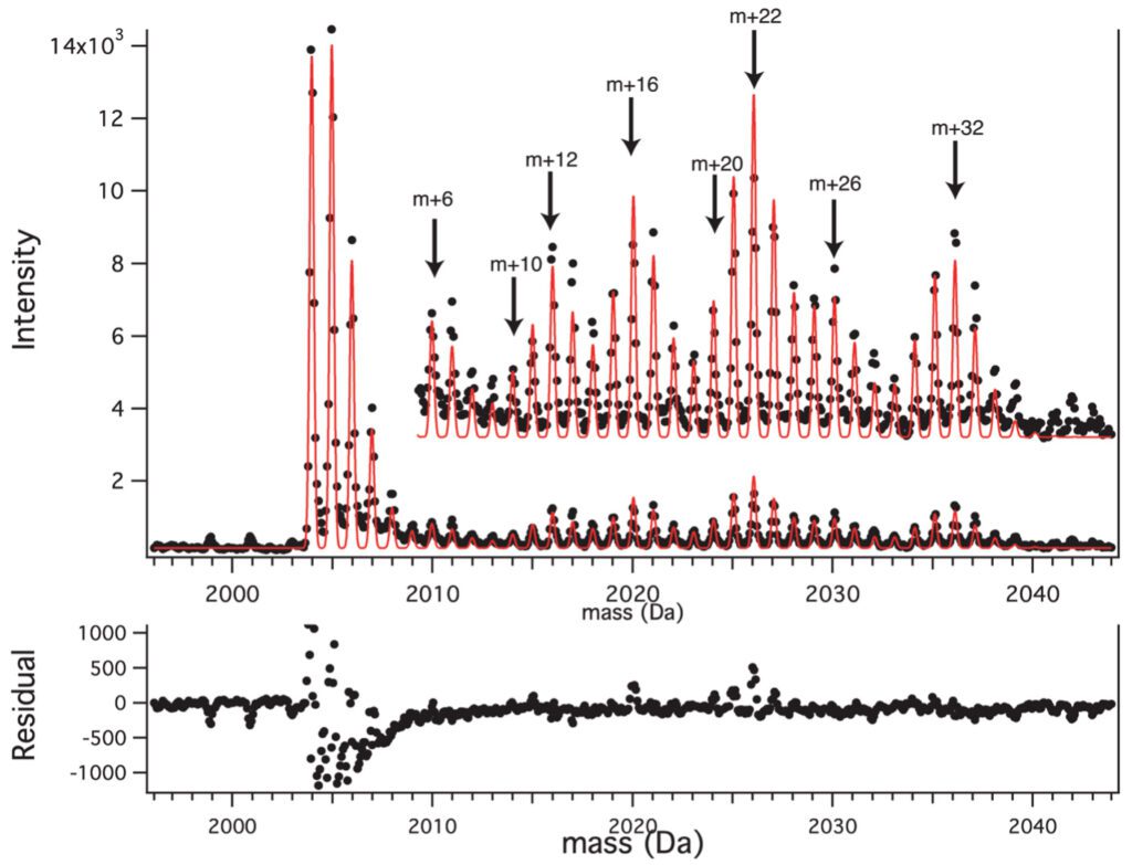
Abstract: Quantitative proteomic mass spectrometry involves comparison of the amplitudes of peaks resulting from different isotope labeling patterns, including fractional atomic labeling and fractional residue labeling. We have developed a general and flexible analytical treatment of the complex isotope distributions that arise in these experiments, using Fourier transform convolution to calculate labeled isotope distributions and least-squares for quantitative comparison with experimental peaks. The degree of fractional atomic and fractional residue labeling can be determined from experimental peaks at the same time as the integrated intensity of all of the isotopomers in the isotope distribution. The approach is illustrated using data with fractional (15)N-labeling and fractional (13)C-isoleucine labeling. The least-squares Fourier transform convolution approach can be applied to many types of quantitative proteomic data, including data from stable isotope labeling by amino acids in cell culture and pulse labeling experiments.
Envelope: Interactive Software for Modeling and Fitting Complex Isotope Distributions
BMC Bioinformatics 2008, 9, 446-455.
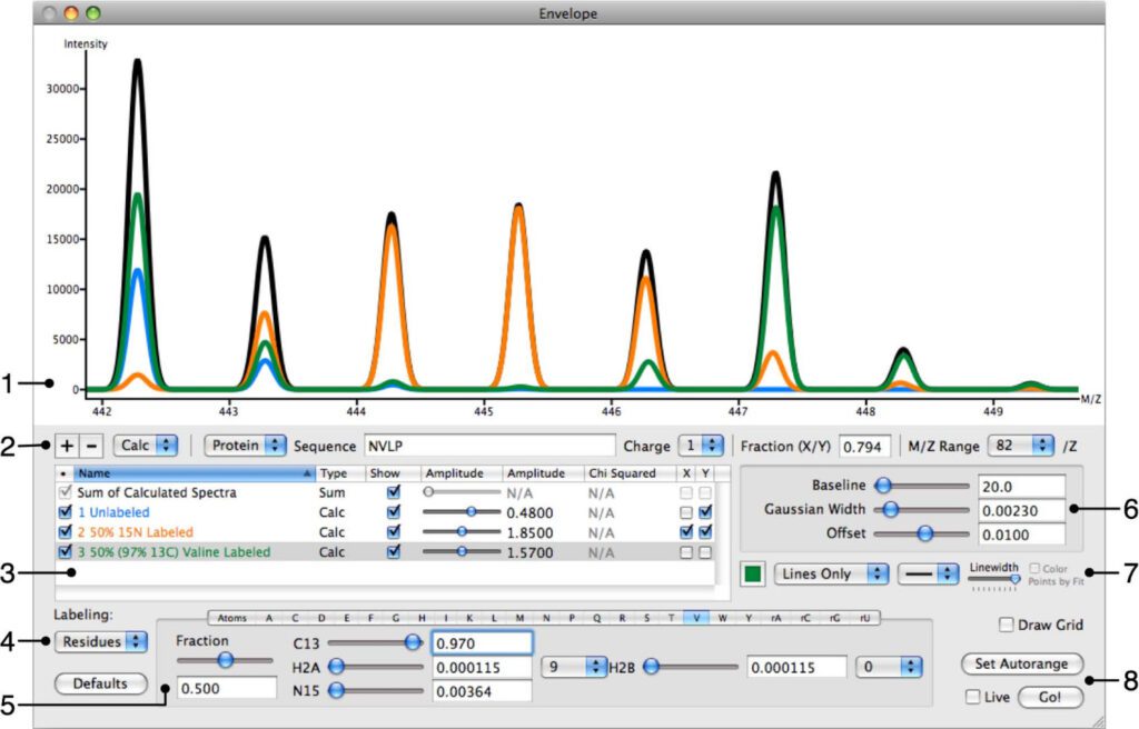
Abstract: An important aspect of proteomic mass spectrometry involves quantifying and interpreting the isotope distributions arising from mixtures of macromolecules with different isotope labeling patterns. These patterns can be quite complex, in particular with in vivo metabolic labeling experiments producing fractional atomic labeling or fractional residue labeling of peptides or other macromolecules. In general, it can be difficult to distinguish the contributions of species with different labeling patterns to an experimental spectrum and difficult to calculate a theoretical isotope distribution to fit such data. There is a need for interactive and user-friendly software that can calculate and fit the entire isotope distribution of a complex mixture while comparing these calculations with experimental data and extracting the contributions from the differently labeled species. Envelope has been developed to be user-friendly while still being as flexible and powerful as possible. Envelope can simultaneously calculate the isotope distributions for any number of different labeling patterns for a given peptide or oligonucleotide, while automatically summing these into a single overall isotope distribution. Envelope can handle fractional or complete atom or residue-based labeling, and the contribution from each different user-defined labeling pattern is clearly illustrated in the interactive display and is individually adjustable. At present, Envelope supports labeling with 2H, 13C, and 15N, and supports adjustments for baseline correction, an instrument accuracy offset in the m/z domain, and peak width. Furthermore, Envelope can display experimental data superimposed on calculated isotope distributions, and calculate a least-squares goodness of fit between the two. All of this information is displayed on the screen in a single graphical user interface. Envelope supports high-quality output of experimental and calculated distributions in PNG or PDF format. Beyond simply comparing calculated distributions to experimental data, Envelope is useful for planning or designing metabolic labeling experiments, by visualizing hypothetical isotope distributions in order to evaluate the feasibility of a labeling strategy. Envelope is also useful as a teaching tool, with its real-time display capabilities providing a straightforward way to illustrate the key variable factors that contribute to an observed isotope distribution. Envelope is a powerful tool for the interactive calculation and visualization of complex isotope distributions for comparison to experimental data.
Improvement in the Apparent Mass Resolution of Oligonucleotides by Using 12C/14N-Enriched Samples
Analytical Chem. 2002, 74, 226-231.
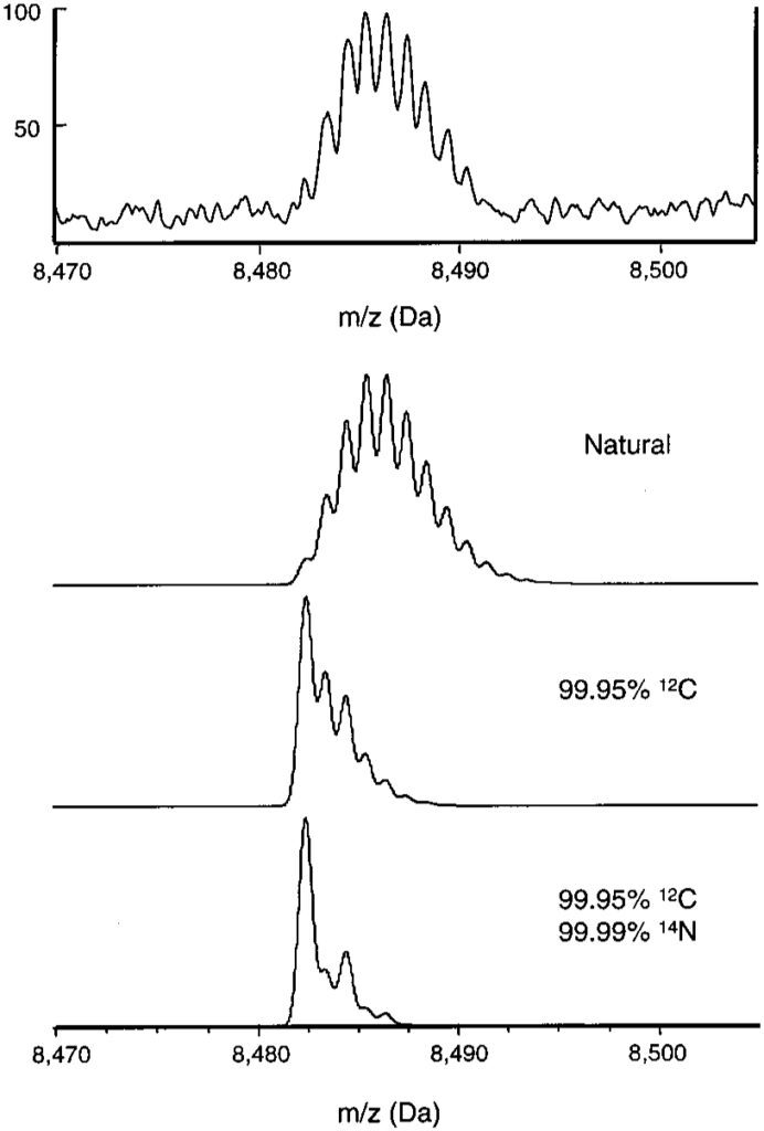
Abstract: The apparent mass resolution of oligonucleotides in time-of-flight (TOF) mass spectrometers has been examined. In a reflectron TOF instrument, where the isotopic profile can be completely resolved, the apparent resolution matches the instrument’s resolving power. In a linear TOF instrument, unresolved isotopic profiles limit the apparent resolution to much lower values than the actual instrument resolution. By using 12C/14N-enriched oligonucleotides, the apparent resolution can be improved significantly. The isotope enrichment method also enhances the signal-to-noise ratio.
Products and Services are for Laboratory Research Applications Only — not for drug, diagnostic, household, or other purposes. Persons intending to use these products and services for other applications do so at their own responsibility and must comply with all appropriate regulations.